3) Implantation
No detailed studies on ailurid implantation are known to me, and Mossman (1987) stated that no panda placenta of the lesser panda had been described. There are a number of descriptions of raccoon placentas, however. Their placenta is most closely of an endothelio-chorial character, although a "vaso-chorial relation has been suggested to be a better name. That is because there is a prominent basement membrane of the fetal sinusoids. The placenta of raccoons falls into the general arrangement of carnivore placentas (Amoroso, 1961).
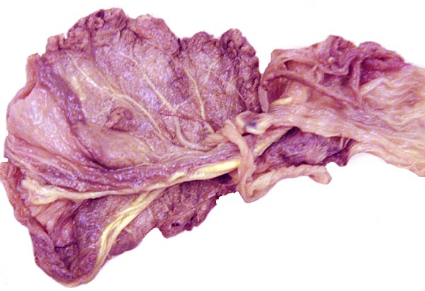
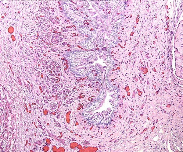
The placenta of the red panda is also very different from the placenta of dog and cat (see those chapters). The filiform arrangement of villi, for instance, is lacking and the placenta does not have the ring-shaped, zonary form seen in many other carnivore species. It is rather discoid with a small central sulcus connecting the villous tissue with a strip of membranes and, in its histology, it also appears to me more similar to the placenta of the primitive carnivore Zorilla striata, described by Rau (1925). A nearly bidiscoid (rather than zonary) placenta was found in that species, and the lesser panda placenta is otherwise also not too much different in structure. The hypertrophy of maternal capillary endothelium and gross morphology are nearly identical to all the carnivore species placentas but they are quite different from those of cats. It must be emphasized, however, that it will be essential to witness an implanted placenta in order to be able to rule out a more ring-shaped organ than that here portrayed. In further consideration of this morphology it is interesting to note that some genetic studies have also likened the lesser panda to mustelids (see above).
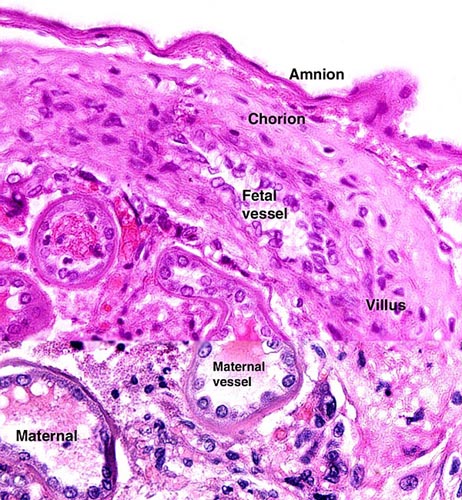
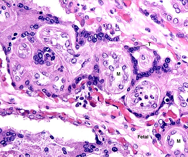
It is noted that possible yolk sac vessels are seen in the cord but no remnant of yolk sac morphology is identified in the specimen studied by me.
4) General Characterization of the Placenta
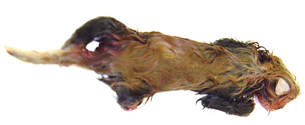
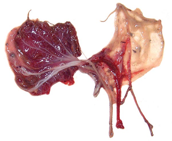
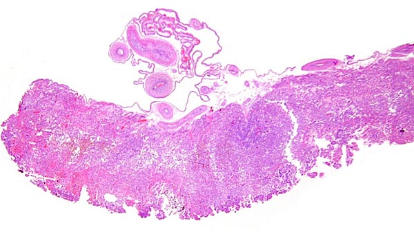
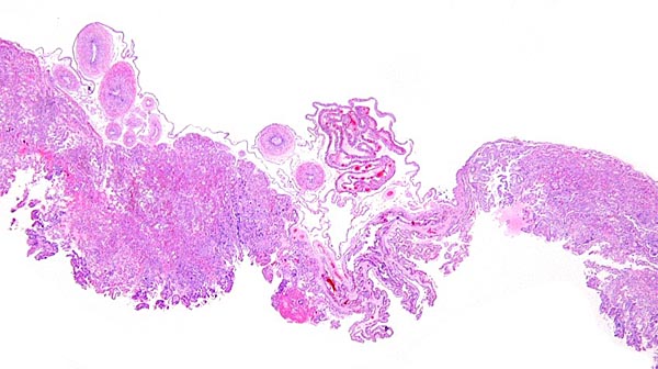
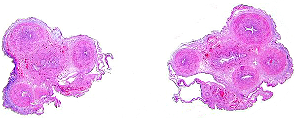
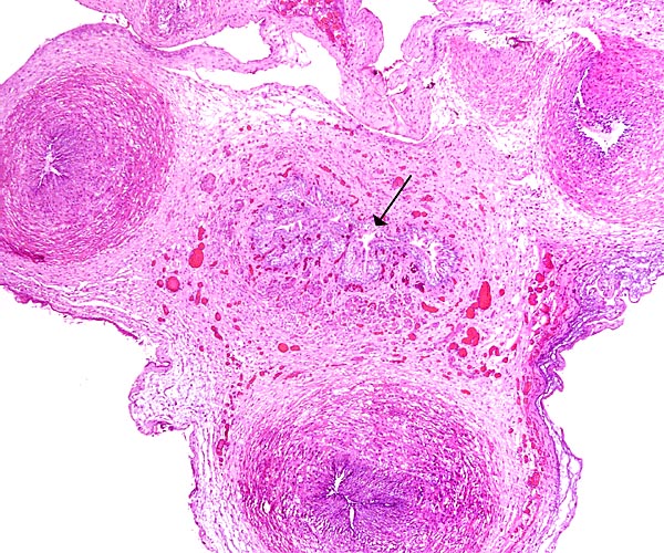
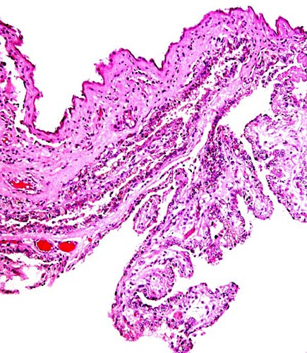
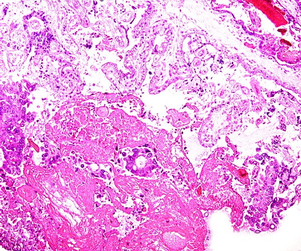
As indicated earlier, I have been unable to find any record of a red panda placenta in the literature. This may thus be its first description. This placenta was detached from the surviving neonate very shortly after its birth and was thus well preserved. It is not known whether the mother consumed any of it, although the organ appeared to be complete. It weighed 5.9 g and measured 4.5 x 3.5 cm, with a 3.5 cm umbilical cord and torn membranes. It had a discoid shape, unlike than the completely zonary placenta of raccoons, dogs and cats. There also was no green discoloration at the edge, as is seen in dog and cat, a feature whose lack was already remarked upon for the raccoon placenta by Watson (1881). Biggers & Creed (1962) were critical of Watson's study and subsequently obtained much better material for study. They mention for instance, that although affirming the zonary nature of the placenta (being certain that it was not interrupted), they were certain that the "epitrichium" described by Watson was actually the closely-applied amnion. Moreover, they were the first to introduce the notion of the "haemophagous organ". It exists prominently in young gestations but gradually involutes towards term. Here, trophoblast is believed to invade the uterus, thus creating focally a hemochorial organ and depositing much pigment. In later studies (Creed & Biggers, 1963; 1964) the authors identified similar structures in several mustelidae and suggested that it is used for red cell phagocytosis of the fetus. These species also had completely annular placentas.
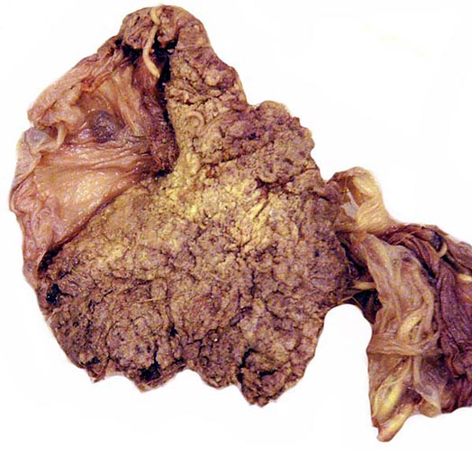
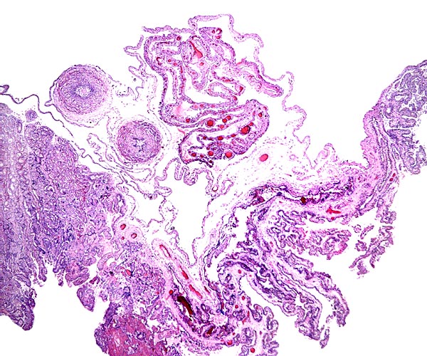
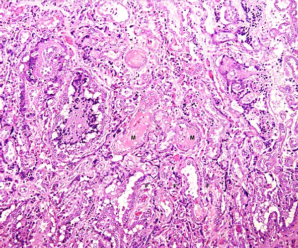
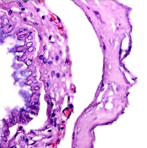
The delivered red panda placenta was 2-3 mm thick and the maternal surface was ragged without any recognizable maternal components. It is noteworthy that the raccoon placenta, as already described in Watson's remarkable paper (1881), was zonary - "The foetus was encircled by an annular or zonary placenta…". The same author, however, also mentions the existence of a "gap" in the placenta and that this "gap" is much more pronounced in mustelids. Thus, the discoid feature of the lesser panda placenta separates the structure from raccoons and certainly from cat and dog, and perhaps the gross morphology allies it more closely to that of mustelids. This is questionable only because some of these are described as being zonary as well and as having a haemophagous organ in young gestations of which there was no direct evidence in this term placenta.
In this delivered placenta, the surface vessels were very prominent. Some other vessels that attached to the membranes are not truly identified. They may be the remains of vitelline vessels, as existing in some mustelidae (e.g. the wolverine - see Wislockie & Amoroso, 1956) but I am not certain of this.
The lesser panda placenta was a delivered, detached organ, much like the detached zonary raccoon placenta described by Watson (1881). He then already remarked on the "deciduate" nature of the placenta; that is to say, a portion of maternal tissue is shed with detachment, as is the case in other carnivora. In our case, the maternal tissue most easily recognized as having been shed are the large number of maternal vascular tubes shown in more detail in the histologic description, the "labyrinth". As was true of Watson in the raccoon placenta, I was unable to identify other maternal (decidua-like or glandular) tissues in the detached placenta. It must be emphasized though that the histologic structure is of course very different from that of the artiodactyl placenta where the fetal placental villi are dislodged from their caruncular tissue recesses, and very little maternal tissue remains with the delivered placenta.
|
Reddacliff et al. (1993) have described a mosaic animal (35,X/36,XY) with equal numbers of lymphocytes of both types. The animal had been suspected of being "intersexual", was small for its age, appeared immunocompromised, and had a normally bicornuate uterus that reminded the authors of that of a cat. The ovaries were largely composed of undefined cells and tubules but had a portion with oocytes and even developing follicles.
14) Immunology
I am not aware of any definitive studies in this species.
15) Pathological features
A number of diseases are known to afflict red pandas. The best review of a variety of conditions is that by Montali et al. (1984) who reviewed the causes of death of 52 animals at the National Zoo in Washington, DC. Surprisingly, there were three congenital anomalies (anencephaly, truncus arteriosus and hypomelia). Infectious diseases, mostly respiratory, were the principal causes of death in this collection as well as reported from other zoos. In addition, however, there were many deaths (25) of juveniles. Other authors have remarked on high perinatal mortality rates and difficulties in raising youngsters. Various parasitic afflictions and two tumors (mastocytoma and leukemia) were described as well. It is also important to point to the number of reports that show distemper mortality following vaccination. Apparently pandas are quite sensitive to the modified virus. Their sensitivity to heat and humidity were also stressed by the authors.
Various additional pathologic conditions have been reported. Thus, Langan et al. (2000) described a nine-year old panda as having died from Tyzzer's disease (intestinal infection with Clostridium piliforme). Kearns et al. (1999) found skin lesions due to Microsporum gypseum in many captive animals. They responded to antifungal therapy. A fatal abscess reported by Dyer et al. (2000) grew only Chromobacterium violaceum, a common soil organism. Poelma (1975) found infection with Pneumocystis carinii in many animals, including red pandas, and Griner (1983) found tuberculosis in a recently imported animal.
16) Physiologic data
I have not been able to find any significant physiological studies to have been carried out in red pandas. Schweigert et al. (1990) examined the transport of vitamin A in carnivores, including the lesser panda. It was found that circulating vitamin A is bound to lipoproteins and reaches levels in carnivores that, in other mammals would be toxic. The study on bile acid composition by Hagey et al (1993) has been referred to at the beginning of this chapter.
17) Other resources
Cell lines of many lesser pandas and of both subspecies are available from CRES at the Zoological Society of San Diego by contacting Dr. Oliver Ryder at oryder@ucsd.edu.
18) Other remarks - What additional Information is needed?
I quite expect that additional studies will be published to further define whether the lesser panda is a procyonid or more of a mustelid. Perhaps it deserves its own status in the classification of animals. Since this is the first description of its placenta one can only hope that in the future an implanted placenta will be described. Also, is there any evidence of delayed implantation? Much more endocrine work is needed.
Acknowledgement
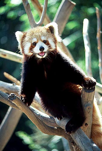
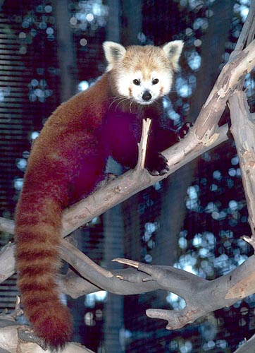
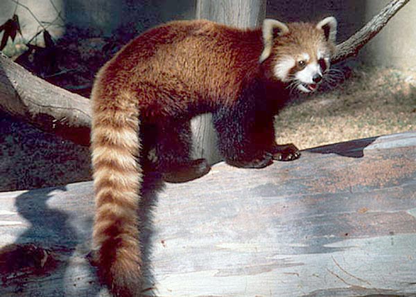
The animal photographs in this chapter come from the Zoological Society of San Diego. I appreciate also very much the help of the pathologists at the San Diego Zoo.
References
Amoroso, E.C.: Placentation. Chapter 15, pp.127-311, In, Marshall's Physiology of Reproduction, V.II, A.S. Parkes, ed. Second Edition. Little, Brown & Co. Boston, 1961.
Biggers, J.D. and Creed, R.F.S.: Two morphological types of placenta in the raccoon. Nature 194:103-105, 1962.
Creed, R.F.S. and Biggers, J.D.: Development of the raccoon placenta. Amer. J. Anat. 113:417-445, 1963.
Creed, R.F.S. and Biggers, J.D.: Placental haemophagous organs in the procyonidae and mustelidae. J. Reprod. Fertil. 8:133-137, 1964.
Creed, R.F.S. and Harrison, R.J.: Preliminary observations on the ultrastructure of the raccoon (Procyon lotor) placenta. J. Anat. 99:933, 1965. {Abstract only}
Dyer, N.W., Krogh, D.F., DeVold, R., Wilson, S.L. and White, D.G.: Chromobacteriosis in a Chinese red panda (Ailurus fulgens styani). J. Vet. Diagn. Invest. 12:177-179, 2000.
Flynn, J.J. and Nedbal, M.A.: Phylogeny of the Carnivora (Mammalia): congruence vs. incompatibility among multiple data sets. Mol. Phylogenet. Evol. 9:414-426, 1998.
Flynn, J.J., Nedbal, M.A., Dragoo, J.W. and Honeycott, R.L.: Whence the panda? Mol. Phylogenet. Evol. 17:190-199, 2000.
Glatston, A.R., ed.: Red Panda Biology. SPB Academic Publishing, Amsterdam, 1989.
Griner, L.A.: Pathology of Zoo Animals. Zoological Society of San Diego, San Diego, California, 1983.
Hagey, L.R., Crombie, D.L., Espinosa, E., Carey, M.C., Igimi, H. and Hofmann, A.F.: Ursodeoxycholic acid in the ursidae: biliary bile acids of bears, pandas, and related carnivores. J. Lipid Res. 34:1911-1917, 1993.
Kearns, K.S., Pollock, C.G. and Ramsay, E.C.: Dermatophytosis in red pandas (Ailurus fulgens fulgens): a review of 14 cases. J. Zoo Wildl. Med. 30:561-563, 1999.
Langan, J., Bemis, D., Harbo, S., Pollock, C. and Schumacher, J.: Tyzzer's disease in a red panda (Ailurus fulgens fulgens). J. Zoo Wildl. Med. 31:558-562, 2000.
Mayr, E.: Uncertainty in science: is the giant panda a bear or a raccoon? Nature 323:769-771, 1986.
Montali, R.J., Roberts, M., Freeman, R.A. and Bush, M.: Pathology survey of the red panda (Ailurus fulgens). Chapter 13 (pp. 128-140) in, "One Medicine", O.A. Ryder and M.L. Byrd, eds. Springer-Verlag, New York, 1984.
Mossman, H.W.: Vertebrate Fetal Membranes. MacMillan, Houndmills, 1987.
Mossman, H.W. and Duke, K.L.: Comparative Morphology of the Mammalian Ovary. University of Wisconsin Press, Madison, Wisconsin, 1973.
Nie, W., Wang, J., O'Brien, P.C., Fu, B., Ferguson-Smith, M.A. and Yang, F.: The genome phylogeny of domestic cat, red panda and five mustelid species revealed by comparative chromosome painting and G-banding. Chromosome Res. 10:209-222, 2002.
Pagel, T.: Der kleine Panda (Ailurus fulgens) - Haltung und Zucht im Zoologischen Garten Köln. Z. des Kölner Zoo 39:139-155, 1996. (In German)
Poelma, F.G.: Pneumocystis carinii infections in zoo animals. Z. Parasitenk. 46:61-68, 1975.
Princée, F.P.G.: Genetic variation in the zoo population of the red panda subspecies Ailurus fulgens fulgens. Zoo Biol. 7:219-231, 1988.
Puschmann, W.: Zootierhaltung. Vol. 2, Säugetiere. VEB Deutscher Landwirtschaftsverlag Berlin, 1989.
Rau, A.S.: Contributions to our knowledge of the structure of the placenta of mustelidae, ursidae, and sciuridae. Proc. Zool. Soc. London 25:1027-1070, 1925.
Reddacliff, G.L., Halnan, C.R.E. and Martin, I.C.A.: Mosaic 35,X/36,XY karyotype and intersex in a red panda (Ailurus fulgens). J. Wildl. Dis. 29:169-173, 1993.
Roberts, M.: The red panda: its history and fragile hold on the future. Your Cincinnati Zoo News Spring/Summer 1983, pp. 1-5.
Roberts, M. and Kessler, D.S.: Reproduction in red pandas, Ailurus fulgens (Carnivora: Ailuropodidae). J. Zool. Lond. 188:235-249, 1979.
Schweigert, F.J., Ryder, O.A., Rambeck, W.A. and Zucker, H.: The majority of vitamin A is transported as retinyl esters in the blood of most carnivores. Comp. Biochem. Physiol. 95A:573-578, 1990.
Slattery, J.P. and O'Brien, S.J.: Molecular phylogeny of the red panda (Ailurus fulgens). J. Hered. 86:413-422, 1995.
Spanner, A., Stone, G.M. and Schultz, D.: Excretion profiles of some reproductive steroids in the faeces of captive Nepalese red panda (Ailurus fulgens fulgens). Reprod. Fertil. 9:565-570, 1997.
Su, B., Fu, Y., Wang, Y., Jin, L. and Chakraborty, R.: Genetic diversity and population history of the red panda (Ailurus fulgens) as inferred from mitochondrial; DNA sequence variation. Mol. Biol. Evol. 18:1070-1076, 2001.
Tagle, D.A., Miyamoto, M.M., Goodman, M., Hofmann, O., Braunitzer, G., Göltenboth, R. and Jalanka, H.: Hemoglobin of pandas: Phylogenetic relationships of carnivores as ascertained with protein sequence data. Naturwissenschaften 73:512-514, 1986.
Tian, Y., Nie, W.H., Wang, J.H., Yang, Y.F. and Yang, F.T.: Comparative chromosome painting shows the red panda (Ailurus fulgens) has a highly conserved karyotype. Yi Chuan Xue Bao 29:124-127, 2002. (In Chinese)
Todd, N.B. and Pressman, S.R.: The karyotype of the lesser panda (Ailurus fulgens) and general remarks on the phylogeny and affinities of the panda. Carnivore Genet Newsl. 5:105, 1968.
Watson, M.: On the female organs and placentation of the raccoon (Procyon lotor) Proc. Roy. Soc. London 32:272-298, 1881.
Wislockie, G.B. and Amoroso, E.C.: The placenta of the wolverine (Gulo gulo luscus) (Linnaeus). Bull. Mus. Compar. Zool. (Harvard). 114:93-100, 1956.
Wurster-Hill, D.H. and Gray, C.W.: The interrelationships of chromosome banding patterns in procyonids, viverrids, and felids. Cytogenet. Cell Genet. 15:306-331, 1975.
Zhang, Ya-Ping and Ryder, O.A.: Mitochondrial DNA sequence evolution in the arctoidea. Proc. Natl. Acad. Sci. 90:9557-9561, 1993.
|