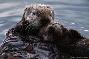
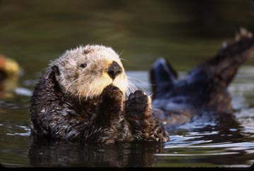
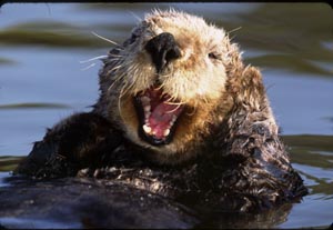
Sea OtterEnhydra lutrisOrder: Carnivora |
||||||||||||||||||||||||||||||
|
||||||||||||||||||||||||||||||
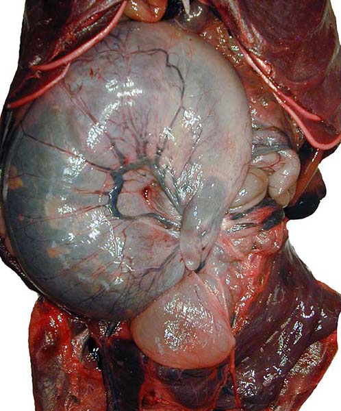
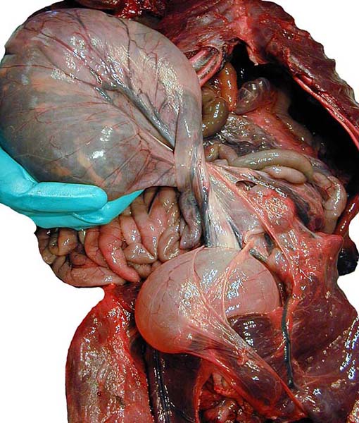
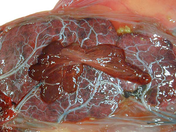
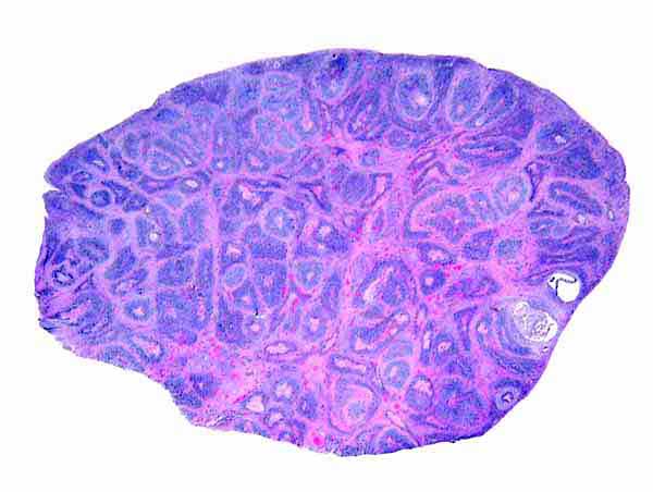
| 2) General Gestational Data Nowak (1999) states that 2% of births are multiple but that the female can successfully raise only one pup. Newborns weigh 1.4-2.3 kg and the estimated length of gestation is 4-5.5 months with some delayed implantation being likely. Antrim & Cornell (1980) summarized the poor reproductive successes of sea otters in captivity. Few animals have survived to maturity when they were raised in captivity. This is contrary to the reproductive rate of increase (15-18%) in the undisturbed wild (Estes et al., 1996). Antrim & Cornell (1980) also commented on the frequency of stillbirths and abortions in first pregnancies and suggested that the preimplantation stage may last 7-8 months, with the implantation stage being 4½-5 months. Jameson & Johnson (1993) believe that the preimplantation period is only 2-3 months long. Larson et al. (2003) indicated that the implanted gestation lasts on average 217 days. It is still unknown, however, whether the ovulation is induced. 3) Implantation This is a labyrinthine, endotheliochorial placenta, quite similar to that of the clawless otter whose chapter should be consulted as that placenta was slightly better preserved. This specimen was made available to me through the cooperation of Melissa Miller and Sharon Toy-Choutka of the Marine Wildlife Veterinary Care and Research Center in Santa Cruz and Davis, California. It comes from a near-term pregnant female that died after being injured and strangled by a fishing line at Monterey, California. The pregnant dam weighed 17.3 kg and had a single male fetus near term that weighed 950 g after autopsy and formalin fixation. It was estimated to be 10 days premature as judged by experienced observers and had some meconium particles over its skin. The ring-shaped placenta weighed 250 g and had implanted in the left horn. |
||||||||||||||||||||||||||||||
|
||||||||||||||||||||||||||||||
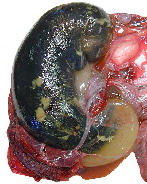
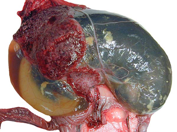
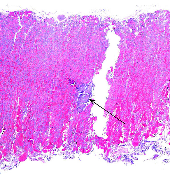
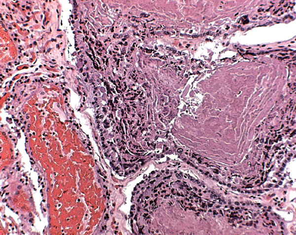
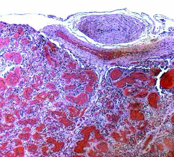
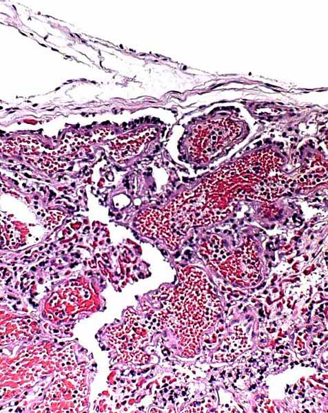
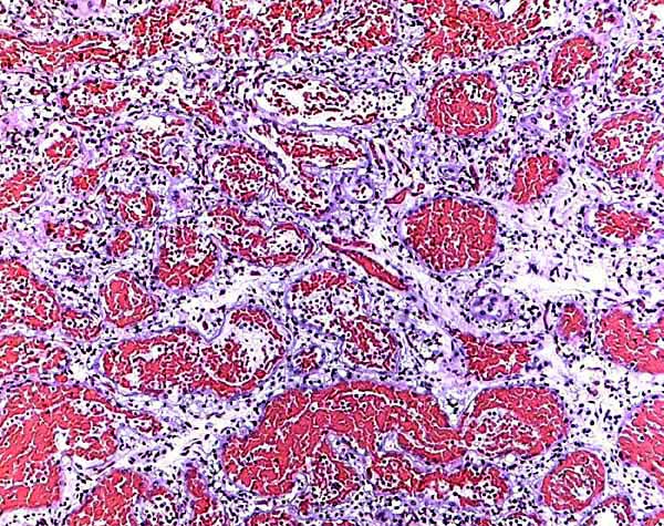
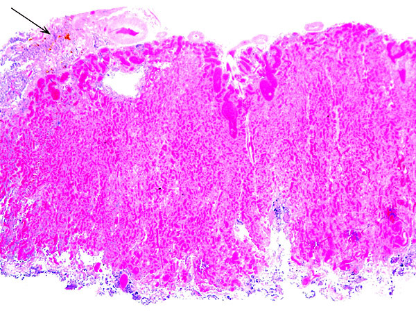
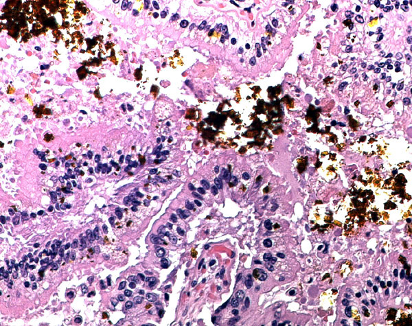
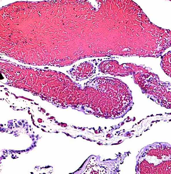
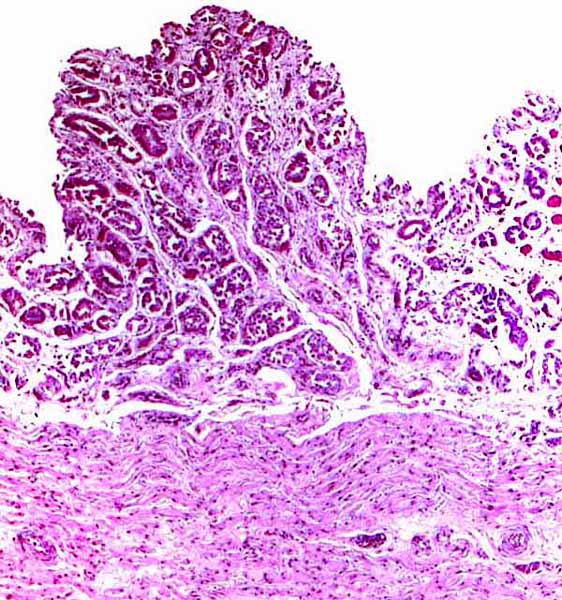
| 5) Details of fetal/maternal barrier This is a lamellar, labyrinthine, endotheliochorial organ similar to that of the clawless otter (see that chapter). The difficult-to-see villi are covered by a single layer of cuboidal trophoblast without any major different cell types being discernible. Amoroso (1961) referred to the trophoblast as "syntrophoblast", but I am not certain that there is truly fusion of trophoblast, and no fine structural detail is available. The villi are composed of very delicate fibrous tissue with distended capillaries. In several sections made from this placenta, only a small hemophagous region was found at the edge of the placenta. It had crystalline pigment in- and outside of trophoblastic cells. As is the usual case with the pigment of the hemophagous organ, this also was negative with Prussian blue stains and was thus not hemosiderin. The box-shaped nature of the yellow crystals suggests hematoidin. In addition though, a saccular structure was present on the fetal surface that has several lobes and contained blood remnants (see above picture). It originates in a central region of modified columnar trophoblast shown next and diagrammed expertly as Fig. 32.1 by Mossman (1987). This lobulated structure was well described and commented upon by Sinha & Mossman (1966) and also in Burton's (1982) review of transplacental iron absorption. It was typically located in the extraembryonic space and also shown to compress the amnionic sac. |
||||||||||||||||||||||||||||||
|
||||||||||||||||||||||||||||||
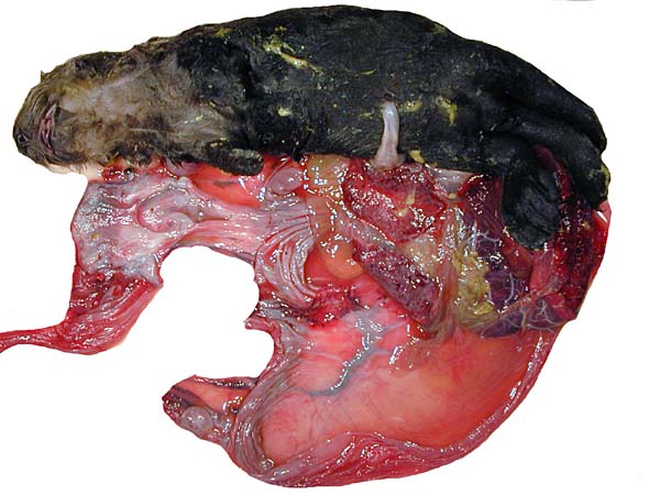
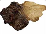
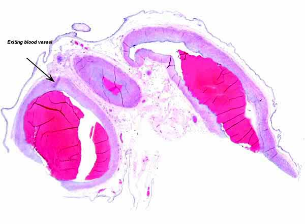
| 6) Umbilical cord As can be seen from the gross photograph, the umbilical cord is extremely short. It has no spirals and the surface is smooth without caruncles. In addition to the three large allantoic vessels, many small blood vessels are present in the umbilical cord, as will be seen in the next photograph. The surface is composed of a single layer of amnion. A typical allantoic duct is present that opens into the complex allantoic sac. |
||||||||||||||||||||||||||||||
|
||||||||||||||||||||||||||||||
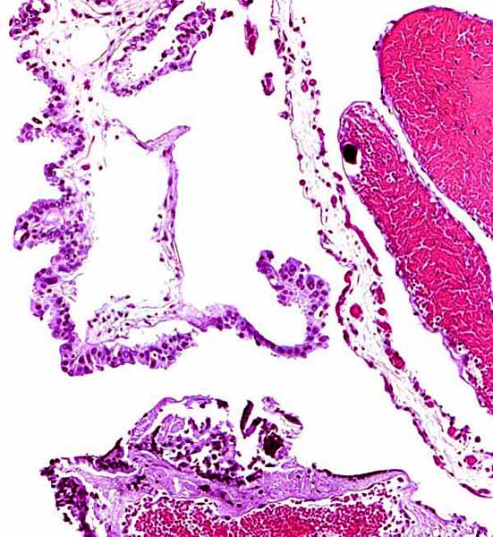
| 7) Uteroplacental circulation There are no published studies. 8) Extraplacental membranes The amnion is avascular and very thin it is composed of a minor amount of connective tissue and has a thin flat surface epithelium. The allantoic sac has a more columnar epithelium and contains finely distributed small blood vessels. The yolk sac, earlier to be found within the umbilical cord, has disappeared at this stage of development. |
||||||||||||||||||||||||||||||
|
||||||||||||||||||||||||||||||
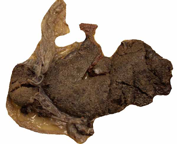
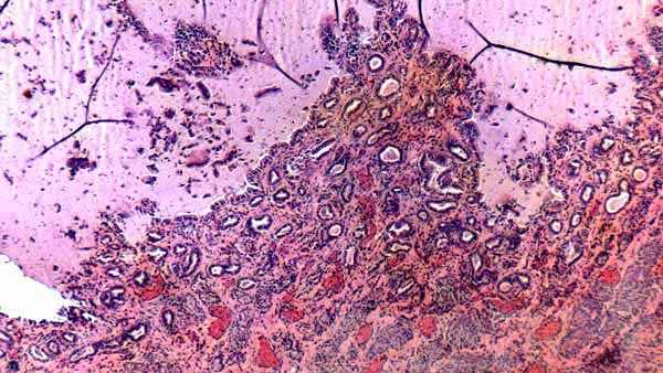
| 9) Trophoblast external to barrier No infiltration of trophoblast into the maternal tissue was seen. 10) Endometrium The uterus of the sea otter has been described as being long and tubular and specially adapted for bearing one or two young (Mossman, 1987). The emptied uterus weighed 170 g after fixation. A girdle-shaped region where the placenta had been attached and which was covered with debris was seen macroscopically. The remainder of the endometrium shows no decidualization and no trophoblastic infiltration was evident. |
||||||||||||||||||||||||||||||
|
||||||||||||||||||||||||||||||
| 11) Various features Neither subplacenta nor metrial glands exist. 12) Endocrinology Larson et al. (2003) validated estrogen and progesterone values of various reproductive states by fecal metabolite monitoring in hopes of improving captive reproductive management. Primary steroid disposition is via feces in sea otters. Females cycled 3-4 times a year with elevation of total estrogens and usually (but not always) followed by rise in progestagen. During the preimplantation stage there was marked elevation of progestagen. There was a dramatic increase in progestagen levels after implantation and thus pregnancy can be reliably diagnosed. A significant fall in total estrogens occurred shortly before parturition. |
||||||||||||||||||||||||||||||
|
||||||||||||||||||||||||||||||
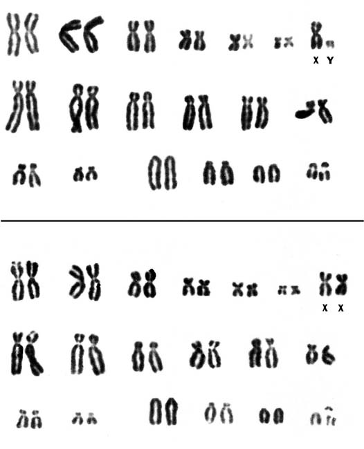
| 13) Genetics Sea otters have 38 chromosomes, the majority being metacentrics (Hsu & Benirschke, 1971). The smallest acrocentric chromosome has the typical "carnivore constriction". Duffield et al. (1996) studied one male and three female animals of a breeding colony and found the male to have several banding abnormalities. They speculated that these may have a causal relation to some congenital defects seen in offspring that were not studied. Hybrids have not been described. |
||||||||||||||||||||||||||||||
|
||||||||||||||||||||||||||||||
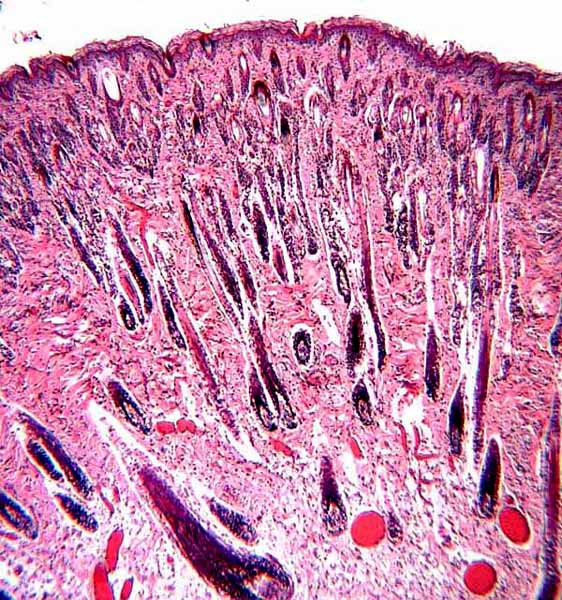
| 14) Immunology King et al. (1996) have cloned and studied the sequence of the IL-6 cDNA fragments of seal, killer whale and sea otter. They found it conserved and the Il-6 of sea otter and seal were closely related to other carnivores, while the IL-6 of killer whale was more similar to artiodactyla. 15) Pathological features There are numerous web sites on sea otters that can be accessed readily by searching in Google under "sea otter". Many contain valuable information. One of the remarkable aspects of sea otters is their skin. Nowak (1999) indicated that this species has the thickest skin, albeit without subcutaneous fat, and that it has extremely dense hair follicles (100,000/sqcm). |
||||||||||||||||||||||||||||||
|
||||||||||||||||||||||||||||||
| 17) Other resources Regrettably no cell lines are available to my knowledge. 18) Other remarks - What additional Information is needed? Acknowledgement Antrim, J.E. and Cornell, L.H.: Reproduction of the Sea otter Enhydra lutris in captivity. Intern. Zoo Yearb. 20:76-80, 1980. Burton, G.J.: Review article. Placental uptake of maternal erythrocytes: a comparative study. Placenta 3: 407-434, 1982. Cornell, L.H., Osborn, K.G., Antrim, J.E. Jr. and Simpson, J.G.: Coccidioidomycosis in a California sea otter (Enhydra lutris). J. Wildl. Dis. 15:373-378, 1979. Dubey, J.R., Rosypal, A.C., Rosenthal, B.M., Thomas, N.J., Lindsay, D.S., Stanek, J.F., Reed, S.M. and Saville, W.J.: Sarcocystis neuronal infections in sea otter (Enhydra lutris): evidence for natural infections with sarcocysts and transmission of infection to opossums (Didelphis virginiana). J. Parasitol. 87:1387-1393, 2001. Dubey, J.P., Zarnke, R., Thomas, N.J., Wong, S.K., van Bonn, W., Briggs, M., Davis, J.W., Ewing, R., Mense, M., Kwok, O.C., Romand, S. and Thulliez, P.: Toxoplasma gondii, Neospora canium, Sarcocystis neurona, and Sarcocystis canis-like infections in marine mammals. Vet. Parasitol. 116:275-296, 2003a. Dubey, J.P., Lindsay, D.S., Rosenthal, B.M. and Thomas, N.J.: Sarcocysts of an unidentified species of Sarcocystis in the sea otter (Enhydra lutris). J. Parasitol. 89:397-399, 2003b. Duffield, D.A., Chamberlin-Lea, J. and Antrim, J.E.: A complex chromosome rearrangement in the karyotype of a wild-caught male sea otter. Endangered Species Update (in The Otter Project) 13 (#12):1-8, 1996. Estes, J.A.: Enhydra lutris. Mammalian Species. Amer. Soc. Mammal. # 133, 1980. Estes, J.A., Doak, D.F., Bodkin, J.R., Jameson, R.J., Monson, D., Watt, J. and Tinker, M.T. Comparative demography of sea otter populations. Endangered Species Update 13:11-13, 1996. Gotch, A.F.: Mammals - Their Latin Names Explained. Blandford Press, Poole, Dorset, 1979. Gulland, F.: Domoic acid toxicity in California sea lions (Zalophus californianus) stranded along the central California coast, May-October 1998. Report to the National Marine Fisheries Service Working Group on unusual Marine Mammal Mortality Events. US Dept. Commerce NOAA Technical Memorandum NMFS-OPR-17, December 2000. Hsu, T.C. and Benirschke, K.: An Atlas of Mammalian Chromosomes. Vol. 6: Folio 285, 1971. Springer-Verlag, N.Y. Jameson, R.J. and Johnson, A.M.: Reproductive characteristics of female sea otters. Marine Mammal Science 9:156-167, 1993. Kim, J.H., Kim, B.H., Kim, J.H., Yoo, M.J. and Kim, D.Y.: Lymphosarcoma in a sea otter (Enhydra lutris). J. Wildl. Dis. 38:616-617, 2002. King, D.P., Schrenzel, M.D., McKnight, M.L., Reidarson, T.H., Hanni, K.D., Stott, J.L. and Ferrick, D.A.: Molecular cloning and sequencing of interleukin 6 cDNA fragments from the harbor seal (Phoca vitulina), killer whale (Orcinus orca), and Southern sea otter (Enhydra lutris nereis). Immunogenetics 43:190-195, 1996. Kreuder, C., Miller, M.A., Jessup, D.A., Lowenstine, L.J., Harris, M.D., Ames, J.A., Carpenter, T.E., Conrad, P.A. and Mazet, J.A.: Patterns of mortality in southern sea otters (Enhydra lutris nereis) from 1998-2001. J. Wildl. Dis. 39:495-509, 2003. Larson, S., Jameson, R., Etnier, M., Fleming, M. and Bentzen, P.: Loss of genetic diversity in sea otters (Enhydra lutris) associated with the fur trade of the 18th and 19th centuries. Mol. Ecol. 11:1899-1903, 2002. Larson, S., Casson, C.J. and Wasser, S.: Noninvasive reproductive steroid hormone estimates from fecal samples of captive female sea otters (Enhydra lutris). Gen. Comp. Endocrinol. 134:18-25, 2003. Lipscomb, T.P., Harris, R.K., Moeller, R.B., Pletcher, J.M., Haebler, R.J. and Ballachey, B.E.: Histopathologic lesions in sea otters exposed to crud oil. Vet. Pathol. 30:1-11, 1993. Miller, M.A., Grigg , M.E. , Kreuder, C., James, E.R., Melli, A.C., Crosbie, P.R., Jessup, D.A., Boothroyd, J.C., Brownstein, D. and Conrad, P.A.: An unusual type of Toxoplasma gondii is common in California sea otters ( Enhydra lutris nereis ) and is a cause of mortality. Int. J. Parasitol. 34:275-284, 2004. Mossman, H.W.: Vertebrate Fetal Membranes. MacMillan, Houndmills, 1987. Nowak, R.M.: Walker's Mammals of the World. 6th ed. The Johns Hopkins Press, Baltimore, 1999. Reimer, D.C. and Lipscomb, T.P.: Malignant seminoma with metastasis and Herpesvirus infection in a free-living sea otter (Enhydra lutris). J. Zoo Wildl. Med. 29:35-39, 1998. Sinha, A.A. and Mossman, H.W.: Placentation of the sea otter. Amer. J. Anat. 119:521-554, 1966. Staveley, C.M., Register, K.B., Miller, M.A., Brockmeier, S.L., Jessup, D.A. and Jang, S.: Molecular and antigenic characterization of Bordotella bronchiseptica isolated from a wild southern sea otter (Enhydra lutris nereis) with severe suppurative bronchopneumonia. J. Vet. Diagn. Invest. 15:570-574, 2003. Stetzer, E., Williams, T.D. and Nightingale, J.W.: Cholangiocellular adenocarcinoma, leiomyoma, and pheochromocytoma in a sea otter. J. Amer. Vet. Med. Assoc. 179:1283-1284, 1981. Thenius, E. and Hofer, H.: Stammesgeschichte der Säugetiere. Springer-Verlag, Berlin, 1960. Williams, T.D. and Pulley, L.T.: Leiomyomas in two sea otters, Enhydra lutris. J. Wildl. Dis. 17:401-404, 1981. |