
Southern White Rhinoceros (or square-lipped rhinoceros)

Ceratotherium simum simum

Order: Perissodactyla
Family: Rhinocerotidae

1) General zoological data of species


There are five species of rhinoceroses, two African and three Asiatic. The Southern white rhinoceros was once seriously endangered but has made a comeback because of conservation measures and translocations. The subspecies of Northern white rhinoceros (C. s. cottoni), on the other hand, is nearly extinct. There is no reason to believe, however, that its placentation differs substantially from the data provided on the Southern subspecies. Zoogeographical and evolutionary data are to be found by Groves (1972, 1975). After serious reduction in numbers of the Southern white rhinoceros, breeding colonies still exist in South Africa and also at the Zoological Society of San Diego, where a herd was assembled in 1972 and large numbers of offspring were produced.

2) General gestational data

Adult white rhinoceroses weigh approximately 3,200-3,600 kg. Their gestational length is 530-550 days. Indian rhinoceroses gestate for about 462-491 days (Laurie et al., 1983). With the exception of size (female black rhinos weigh about 1,600 kg, the Indian rhino weighs 1,600 kg), not any significant difference was found in the morphology of placentas from white, black and Indian rhinoceroses. They all implant in both uterine horns, the fetus being located mostly in one horn. The placenta extends into the other horn. The placental weight at term, including membranes, varies between 3,900 and 7,600 g in our large experience (without the short umbilical cord).

Weight at birth is 48.5 kg, and maturity occurs at 3 years in females. Cows as old as 36 years may still bear young (Groves, 1972). Only one young is born. Newborn Indian rhinos weigh 40-80 kg. (Please see the chapter on Indian rhinoceros).

image-1 
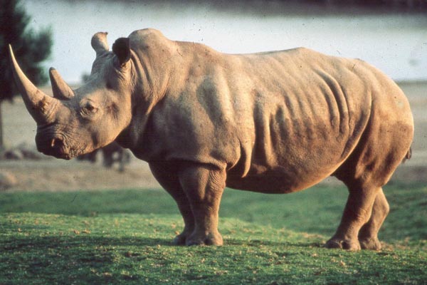
White rhinoceros in the San Diego Zoo's Wild Animal Park.
   

3) Implantation

Early implantational stages were depicted by the sonographic study of Radcliffe et al. (1997). The earliest stage shown occurred 15 days after ovulation, with embryonic definition visible on day 23, and heartbeat visible on day 26.

The exact time of implantation, however, has not yet been defined. The placenta is diffusely villous, with larger streak-like areas of villous absence. This is one reason for the organ to be called "Placenta villosa diffusa incompleta". These bare areas are also referred to as "streets (Strassen)". The organ implants in both uterine horns, with the fetus located mostly in one horn, and the placenta extending to both sides.
 
image-1 
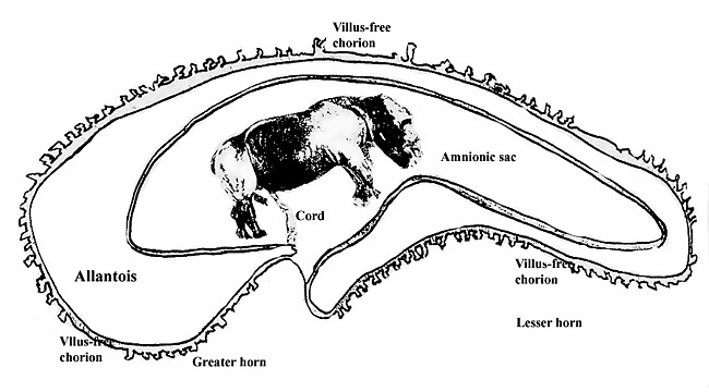
Schematic representation of rhinoceros placentation.
   

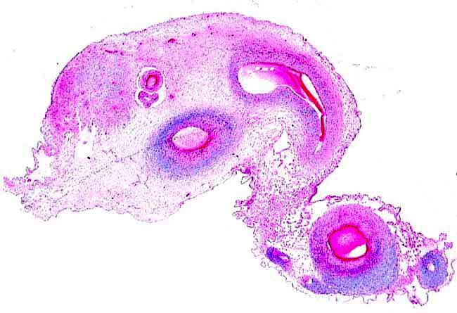
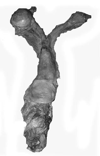
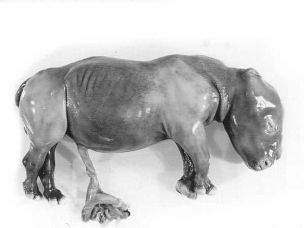
4) General characteristics of placenta

The placentas are very large and very thin. One weighing 7,600 g is depicted here. It measured 190 cm in length, 100 cm in greatest width and 50 cm in smallest diameter. It was 2 mm in thickness. Another placenta examined by me weighed 6,150 g, measured 210x56 cm, was typically thin and had an extremely short cord with four vessels and allantoic duct. The placenta of a term Indian rhinoceros weighed 5,300 g, with an additional 950 g of amnionic and allantoic membranes. It measured 230 cm in greatest width. These are diffuse placenta without cotyledons, and with an epitheliochorial barrier character. Many placentas have on their maternal surfaces band-like connective tissue areas that are bare of villi. They usually follow the lesser curvature of the placenta and have been referred to as "Strassen" (streets) in the Suisse literature.

A very young gestation has recently become available for study. It occurred in an old (35 years or more) female that has had 15 young before and died from intestinal volvulus. The macroscopic pictures are shown next.
 
image-1

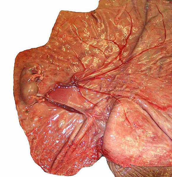
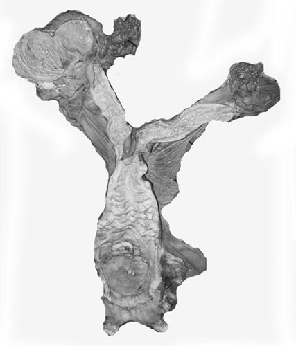
Early stage of white rhino placentation. Embryo is 35 g and is still within the amnion.

   
image-1

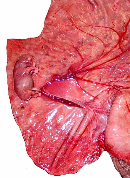
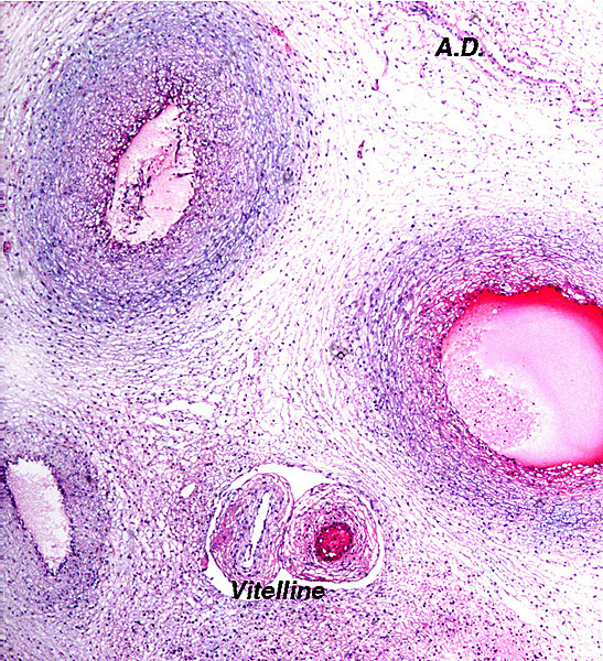
The amnion has been removed and a very short umbilical cord can be seen whose vessels branch into two sections for future occupation of other uterine horn.

   
image-1 
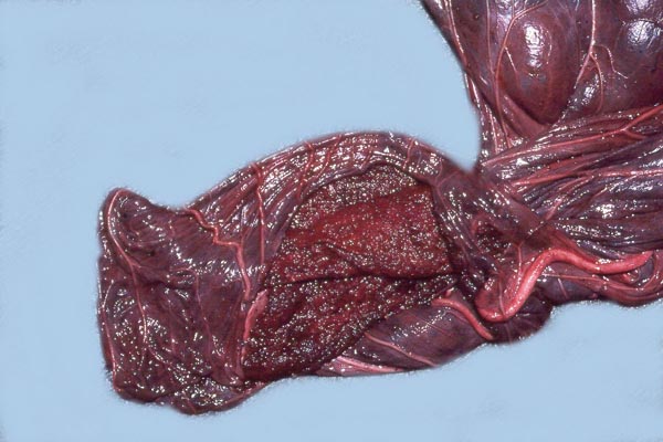
White rhinoceros, delivered placenta, fetal side.
   
image-1 
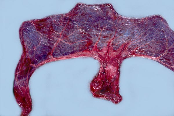
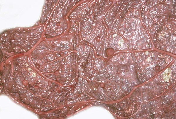
Maternal aspect of white rhinoceros placenta (center).
   

The cord is relatively short and there is a large, heavily vascularized allantoic sac. Hippomanes (yellow/brown) are composed of eosinophilic debris with or without crystalline inclusions (180 g in an Indian rhino). In addition, there are numerous round, yellowish squamous patches projecting on the amnionic surface of some black and Indian rhinoceros placentas; they measure 0.5-1 cm.
 
image-1 Amnionic surface of Indian rhinoceros placenta with round nodules.
   

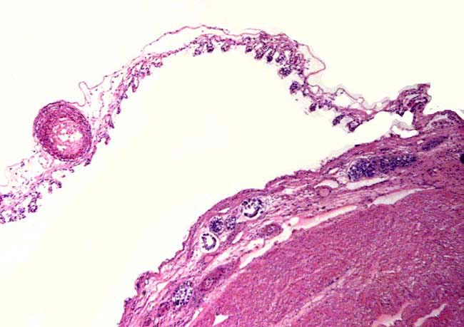
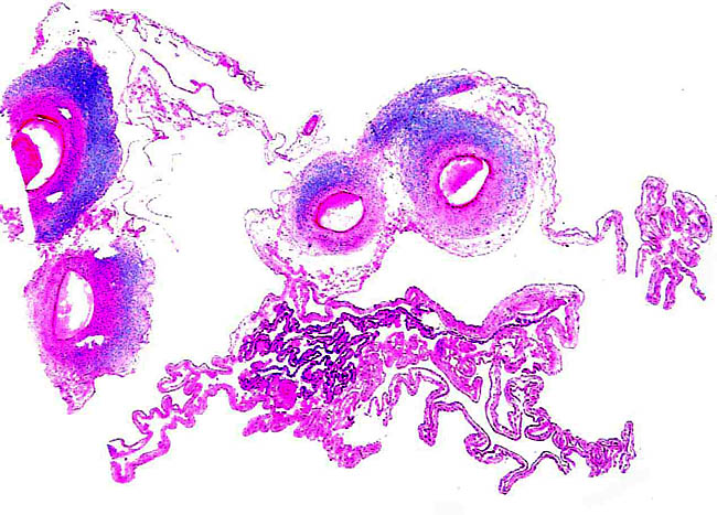
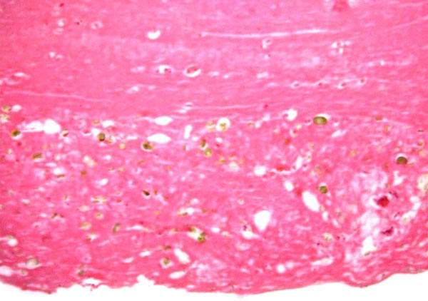
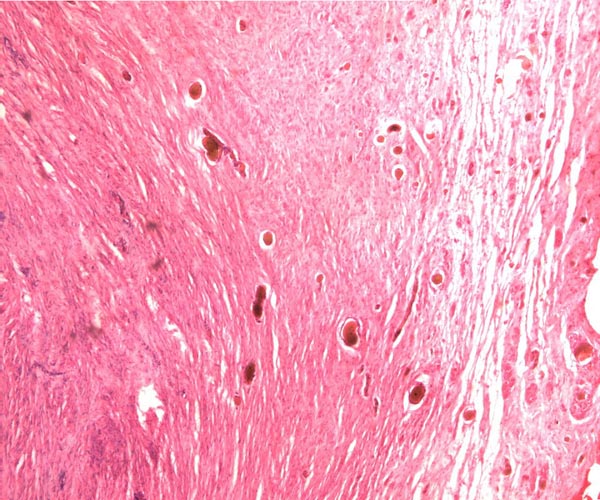
Leaf-like and folded villi were alluded to by Ludwig et al. (1965), representing thinner and others, taller epithelial structures. Histochemical reactions were possible that showed minor differences (Ludwig & Müller, 1965). Occasional binucleated trophoblastic cells have been described, but there is no uterine invasion by trophoblast. The electronmicroscopic study by Ludwig & Villiger (1965) identified similarities to equine placentas; numerous trophoblastic transport vesicles were seen.

The allantoic sac is anchored by thin connective strands to the chorion (Dolinar et al., 1965). While the amnionic epithelium is very thin and flat, the allantoic sac is lined by cuboidal to columnar epithelium. The allantois is diffusely vascularized.
 
image-1 
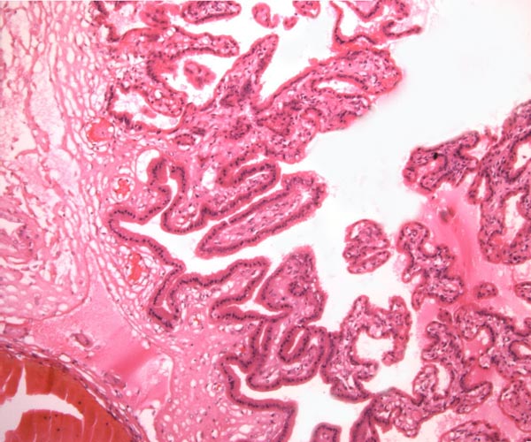
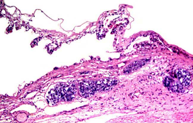
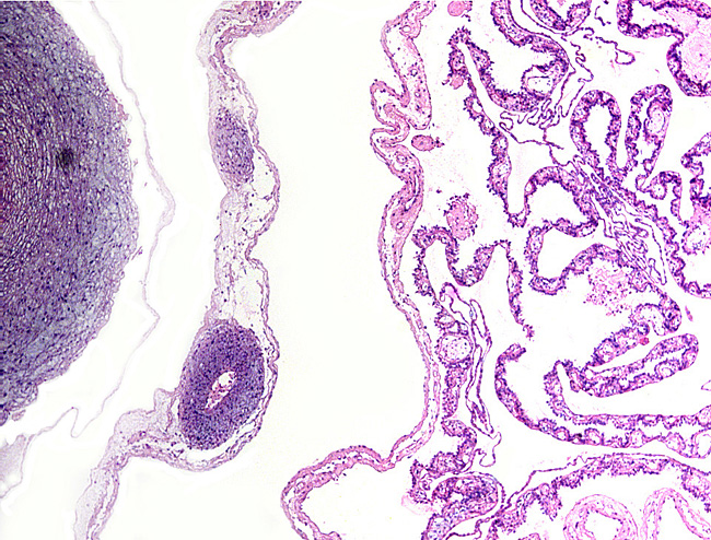
Villi attached to chorion (left) of white rhinoceros.
   
image-1 
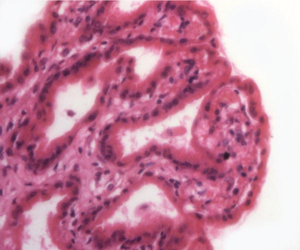
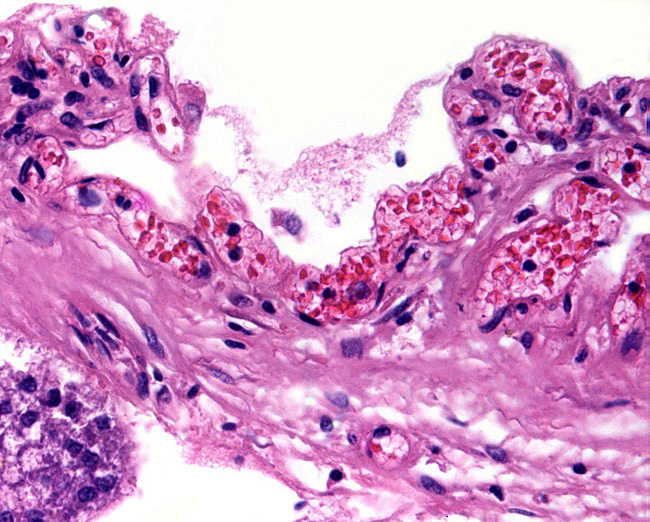
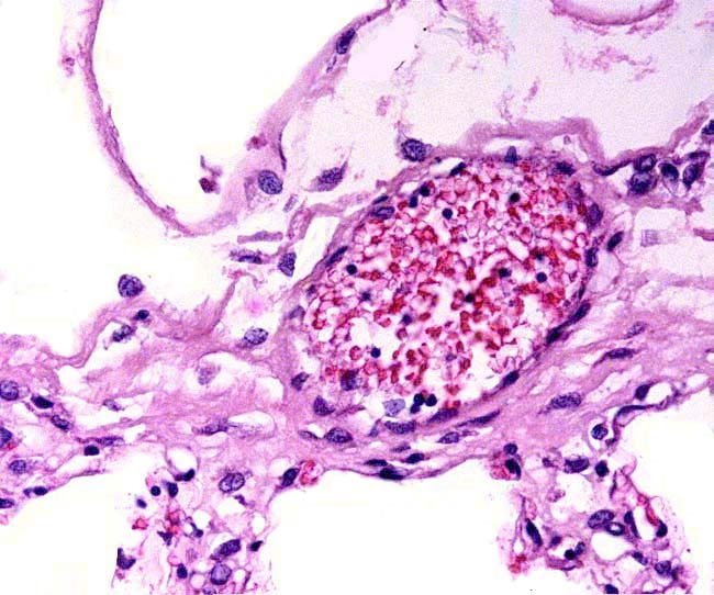
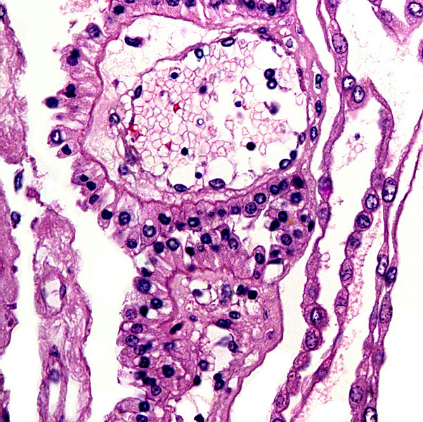
Higher magnification of white rhinoceros villi.


A complete review of all publications on three species of rhinocerotidae placentas was published by Benirschke & Lowenstine (1995). This report contains details in tabular form and is appended, in full, at the end of the chapter on the Indian rhinoceros.

The immature gestation shown above had a 35 g, 72 cm CR male fetus and was in a female whose death was attributed to a twisted colon. She had 15 offspring earlier and was at least 30 years old. The placenta and fetus were in the right horn, the left was not occupied by placenta. It had a polypoid, slightly cystic endometrium. Although autopsy was done within 6 hours of death, considerable autolysis had taken place. The placenta was extremely thin and easily detached from the endometrium. Because of the rarity of such a specimen I attach many pictures of that gestation next.

image-1

Chorionic membrane with fetal vessels attaching to endometrium below.

   
image-1

Higher magnification of attachment. The purple masses are endometrial glands.

   
image-1

The endometrial surface has a vast array of thin capillaries.

   
image-1

This is the attaching placental surface with chorioallantoic blood vessel. Amnion is above, early villi with autolyzed trophoblast below.

   
image-1

Section of membranes directly after the cord has split into two branches. The dark epithelium in the center is presumably the remains of the yolk sac membrane.

   
image-1

Fetal cord vessel at left. Next to it is the amnion and allantoic membrane with allantoic vessels. Membrane at right is the yolk sac remains.

   
image-1

Remains of yolk sac membrane.

   
image-1

Allantoic membrane.

   
image-1 Umbilical cord immediately before its split with yolk sac vessels above left.
   
image-1

Higher magnification of vitelline vessels and allantoic duct (A.D.).

   
image-1

Edematous endometrium of unoccupied uterine horn.


5) Details of barrier structure

This is a typical diffuse epithelial-chorial (appositional) placenta without evidence of invasion of the endometrium.

6) Umbilical cord

The umbilical cord is short, usually with the delivered placenta comes only a 5-6 cm piece, but the portion remaining on the fetus is usually also short and has only one twist. The cord of one of our Indian rhinoceros placentas was 10 cm long. But the cord has not been measured in utero and final measurements are yet to be forthcoming.

 
image-1 
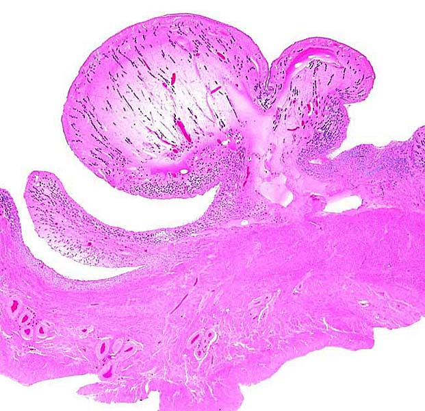
Fetus of Southern white rhinoceros with small portion of placenta attached. Note the short length of the umbilical cord.
   

A remarkably long umbilical cord (57.8 cm) in an Indian rhinoceros was described by Dolinar et al. (1965). The umbilical cord has 4 large umbilical vessels and a very large number of small vessels that provide the circulation for the allantois. It also has a large allantoic duct. A vitelline duct is absent. The surface of umbilical cords has squamous plaques.
 
image-1 Portion of rhinoceros umbilical cord. Large vessel at left; numerous small allantoic vessels are evident.
   

7) Uteroplacental circulation

No descriptions are available.

8) Extraplacental membranes

Only an allantoic sac is present that is diffusely fused to the amnion and, less completely, to the chorion. Free membranes do no exist. Numerous allantoic vessels supply the allantois.

 
image-1 
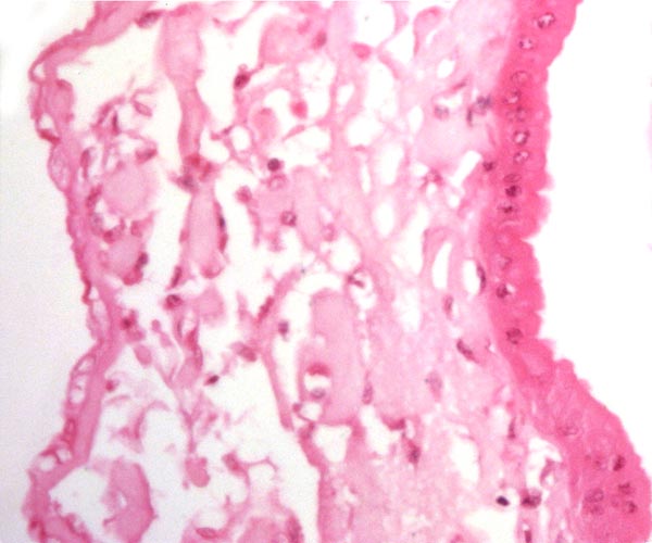
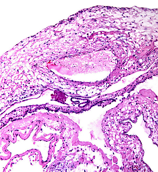
Amnion at left (flat epithelium) allantois right (columnar epithelium).
   

9) Trophoblast external to barrier

There is no trophoblast beyond the villous structures.

10) Endometrium

Decidualization has not been described, but hemosiderin is found in post partum uteri, and many post partum endometria have cystic glands.

11) Various features

The endometrium has hemosiderin-laden macrophages after gestations, suggesting that there is some bleeding during parturition. No implanted placenta has been observed. All those seen by me and those described in the literature had been delivered placentas.

12) Endocrinology

The estrous cycle was defined as being 31-35 days by Radcliffe et al. (1997). This was done in a combined sonographic and endocrine study. Estrus lasts less than 24 hours. Numerous other studies of estrogens and progesterone levels have been performed and are quoted by Miller (1983).

Neonatal gonads display pronounced testicular interstitial cell or ovarian lutein activity, not unlike those of horses. While no gonadotropins have been identified, it is likely that some gonadotropin must present during gestation because of the luteinization of fetal ovaries and stimulation of testicular interstitial cells of neonates. There may thus be similarities to the equine gestation, but hormones have yet to be defined.

Reproductive parameters in zoological gardens have been delineated for the black rhinoceros by Carlstead et al. (1999). A workshop on white rhinoceros steroid determinations and induced ovulation was held in San Diego (Patton et al., 1998) and is available from that institution. Another comprehensive conference volume on rhinoceros biology is available from a 1991 conference (Ryder, 1993).


Patton et al. (1999) monitored reproductive cycles and pregnancy with the assessment of progesterone metabolites from fecal extracts. Cycles lasted more often one or, less commonly, two months. Pregnancy was diagnosed with rising levels at 3 months post-breeding.

The preservation of fecal samples for steroid study has been discussed many times and was controversial when samples had been imported for study. Now, Galama et al. (2004) have published a study that indicates reliable preservation of steroids when stored in methanol or after drying in a solar box cooker. Admittedly, the study was done in black rhinoceros, but should be equally applicable to other species.

13) Genetics

The chromosome number of the white rhinoceros (as well as the black, Indian and Sumatran rhinoceroses) is 2n=82 (although Hansen reported 2n=84). The X-chromosome is the only metacentric element. Many other elements have small short arms, even the Y-chromosome. Three animals of the Northern white rhinoceros had only 81 elements. Mitochondrial DNA was studied in different animals and different subspecies by Ashley et al., (1990), Harley & O'Ryan (1993), and by George et al. (1993). Only the black rhinoceros has 84 chromosomes. Allozyme variation was studied in African and Indian rhinoceroses by Mereniender et al. (1989).

image-1 Male and female karyotypes of white rhinoceros (Hsu & Benirschke, 1973).
   

14) Immunology

No studies are known to us.

15) Pathological features

Veterinary problems, diseases and numerous procedures, were summarized by Silberman & Fulton (1979), and a complete veterinary bibliography was compiled by Miller (1983). It includes references to reproductive laboratory data as well. Dermal problems and infections appear to be the commonest ailments, including pox (Schaller & Pilaski, 1979).

A major problem for black rhinoceroses is the propensity to develop hemolytic anemia (Chaplin, et al., 1986; Miller & Boever, 1982; Paglin et al., 1986).

We have reported huge vacuoles in the trophoblast of a normal placenta from an Indian rhinoceros. This region overlay a region of endometrial degeneration and it does not represent the trophoblastic transport vesicles.

 
image-1 
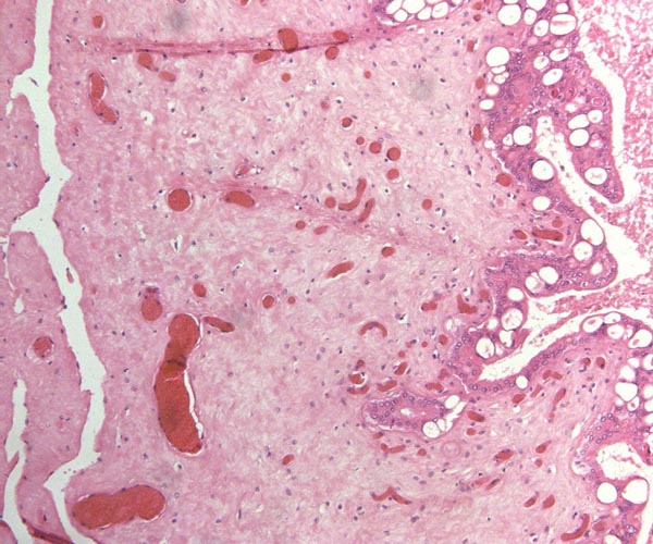
Indian rhinoceros chorionic surface with abnormally vacuolated trophoblast at right, overlying endometrial debris.
   

16) Physiological data

Whatever relevant physiologic data are available have been provided in review form by Silberman & Fulton (1979), and as bibliography by Miller (1983).

17) Other resources

Cell strains of four species of rhinoceros are available from the “Frozen zoo” at the Zoological Society of San Diego by contacting Dr. Oliver Ryder at oryder@ucsd.edu.

None are available of the severely endangered Javan rhinoceros.

References

Ashley, M.V., Melnick, D.J. and Western, D.: Conservation genetics of the black rhinoceros (Diceros bicornis): I. Evidence from the mitochondrial DNA of 3 populations. Conserv. Biol. 1:71-77, 1990.

Benirschke, K. and Lowenstine, L.J.: The placenta of the rhinocerotidae. Verh. Ber. Erkr. Zootiere (Dresden). 37:15-23, 1995.

Carlstead, K., Fraser, J., Bennett, C. and Kleiman, D.G.: Black rhinoceros (Diceros bicornis) in U.S. Zoos: II. Behavior, breeding success, and mortality in relation to housing facilities. Zoo Biol. 18:35-52, 1999.

Chapin, H., Malecek, A.C., Miller, R.E., Bell, C.E., Gray, L.S. and Hunter, V.L.: Acute intravascular hemolytic anemia in the black rhinoceros: Hematologic and immunohematologic observations. Amer. J. Vet. Med. 47:1313-1320, 1986.

Dolinar, Z.J., Ludwig, K.S. und Müller, E.: Ein weiterer Beitrag zur Kenntnis der Placenten der Ordnung Perissodactyla: Zwei Geburtsplacenten des Indischen Panzernashorns. (Rhinoceros unicornis L.). Acta Anat. 61:331-354, 1965.

Galama, W.T., Graham, L.H. and Savage, A.: Comparison of fecal storage methods for steroid analysis in black rhinoceros (Diceros bicornis). Zoo Biol. 23:291-300, 2004.

George, M. Jr., Chemnick, L.G., Cisova, D., Gabrisova, E., Stratil, A. and Ryder, O.A.: Genetic differentiation of white rhinoceros subspecies: diagnostic differences in mitochondrial DNA and serum proteins. In, Proc. Intern. Conference on Rhinoceros Biology and Conservation, San Diego, CA 1991, pp. 105-113.

Groves, C.P.: Taxonomic notes on the white rhinoceros. Ceratotherium simum (Burchell, 1817). Säugetierk. Mitteil. 23:200-212, 1975.

Groves, C.P.: Phylogeny of the living species of Rhinoceros. Z. zool. System. Evol. 21:293-313, 1983.

Hansen, K.M.: Q-bands of some chromosomes of white rhinoceros (Diceros simus). Hereditas 82:205-208, 1976.

Harley, E.H. and O'Ryan, C.: Molecular genetic studies of southern African rhinoceros. In, Proc. Intern. Conference on Rhinoceros Biology and Conservation, San Diego, CA 1991, pp. 101-104.

Heinichen, I.G.: Karyological studies on southern African perissodactyla. Kodoe 13:51-108, 1970.

Houck, M.L., Ryder, O.A., Váhala, J., Kock, R.A. and Oosterhuis, J.E.: Diploid chromosome number and chromosomal variation in the white rhinoceros (Ceratotherium simum). J. Hered. 85:30-34, 1994.

Hsu, T.C. and Benirschke, K.: An Atlas of Mammalian Chromosomes. Vol. 7:Folio 339, 1973. Springer-Verlag, New York.

Hungerford, D.A., Chandra, H.S. and Snyder, R.L.: Somatic chromosomes of a black rhinoceros (Diceros bicornis Gray 1821). Amer. Naturalist 101:357-358, 1967.

Lang, E.M.: Geburt eines Panzernashorns, Rhinoceros unicornis, im Zoologischen Garten Basel. Säugetierk. Mitt. 5:69-70, 1957.

Lang, E.M.: Einige biologische Daten vom Panzernashorn (Rhinoceros unicornis). Rev. Suisse Zool. Genève 74:603-607, 1967.

Laurie, W,A., Lang, E.M. and Groves, C.P.: Rhinoceros unicornis. In, Mammalian Species # 211, pp.1-6, 1983. Amer. Soc. Mammalogy.

Ludwig, K.S.: Zur Kenntnis der Geburtsplacenten der Ordnung Perissodactyla. Acta Anat. 49:154-167, 1962.

Ludwig, K.S. und Villiger, W.: Zur Ultrastruktur der Blattzottenepithelien in der Placenta des Indischen Panzernashorns (Rhinoceros unicornis L.). Acta Anat. 62:593-605, 1965.

Ludwig, K.S. und Müller, E.: Zur Histochemie der Placenta des Panzernashorns (Rhinoceros unicornis L.). Acta Anat. Suppl. 115:155-159, 1965.

Mereniender, A.M., Woodruff, D.S., Ryder, O.A., Kock, R. and Váhala, J.: Allozyme variation and differentiation in African and Indian rhinoceroses. J. Hered. 80:377-382, 1989.

Miller, R.E.: Veterinary Bibliography for Rhinoceros. A.A. Balkema Publ., Amsterdam , 1983.

Miller, R.E. and Boever, W.J.: Fatal hemolytic anemia in the black rhinoceros: Case report and a survey. J.A.V.M.A. 181:1228-1231, 1982.

Morales, J.C., Andau, P.M., Supriatna, J., Zainuddi, Z.Z. and Melnick, D.J.: Mitochondrial DNA variability and conservation genetics of the Sumatran rhinoceros. Conserv. Biol. 11:539-543, 1997.

Paglin, D.E., Valentine, W.N., Miller, R.E., Nakatani, M. and Brockway, R.A.: Acute intravascular hemolysis in the black rhinoceros: Erythrocyte enzymes and metabolic intermediates. Amer. J. Vet. Res. 47:1321-1325, 1986.

Patton, L., Czekala, N. and Lance, V.: Workshop on problems associated with the low rate of reproduction among captive-born female Southern White Rhinoceros (Ceratotherium simum simum). Zoological Society of San Diego, 1998.

Patton, M.L., Swaisgood, R.R., Czekala , N.M. , White, A.M., Fetter, G.A., Montagne, J.P., Rieches, R.G. and Lance, V.A.: Reproductive cycle length and pregnancy in the Southern white rhinoceros (Ceratotherium simum simum) as determined by fecal pregnane analysis and observations of mating behavior. Zoo Biol. 18:111-127, 1999.

Radcliffe, R.W., Czekala, N.M. and Osofsky, S.A.: Technical Report. Combined serial ultrasonography and fecal progestin analysis for reproductive evaluation of the female white rhinoceros (Ceratotherium simum simum): Preliminary results. Zoo Biol. 16:445-456, 1997.

Ramsey, E.: The Placenta of Laboratory Animals and Man. Holt, Rinehart and Winston, NY 1975.

Ryder, O.A., ed.: Rhinoceros Biology and Conservation. Zoological Society of San Diego, 1993 (Proc. of Conference 1991).

Schaller, K. and Pilaski, J.: Pocken bei Breitmaul-nashörnern (Ceratotherium s. simum) im Zoologischen Garten Münster. Zool. Garten 49:169-184, 1979.

Silberman, M.S. and Fulton, R.B.: Medical problems of captive and wild rhinoceros - a review of the literature and personal experiences. J. Zoo Anim. Med. 10:6-16, 1979.

Wurster, D.H. and Benirschke, K.: The chromosomes of the Great Indian Rhinoceros (Rhinoceros unicornis L.). Experientia 24:511, 1968.
Groves, C.P.: Ceratotherium simum. In, Mammalian Species. 8:1-6, 1972. Amer. Soc. Mammalogy.
.
18) Other data to be accumulated

There is a need for the delineation of hormonal profiles during pregnancy because of the fetal gonadal stimulation. The length of umbilical cords is not securely established. No implanted placenta has been observed and thus no definitive statements concerning the fetal/maternal interface can be made.

 
image-1 Hippomanes with crystals in white rhinoceros.
   
image-1 Uterus of black rhinoceros (Courtesy Dr. Montali, Smithsonian Institute, Washington, DC).
   
image-1 Uterus of black rhinoceros (Courtesy Dr. Montali, Smithsonian Institute, Washington, DC).
   

Various Rhinoceroses
 
image-1 
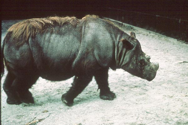
Sumatran rhinoceros at Copenhagen Zoo.
   
image-1 
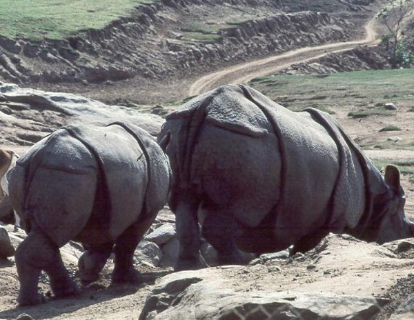
Indian Rhinoceros at San Diego Wild Animal Park.
   
image-1 
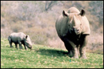
Black rhinoceros with young at San Diego Wild Animal Park.
   
image-1 
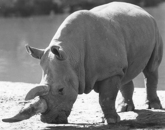
Southern White Rhinoceros at San Diego Wild Animal Park.