Modeling the Human Placenta
While animal models can be used to model some aspects of the human placenta, the unique nature of this organ necessitates development of model systems in which the actual human placental tissue/cells can be studied. Since the majority of placenta-based pregnancy complications arise early in pregnancy, ethically-acceptable models are needed that can recapitulate placental cell types early in gestation. We are currently using three different methods for modeling this unique organ.
Stem cell modeling of placental development.
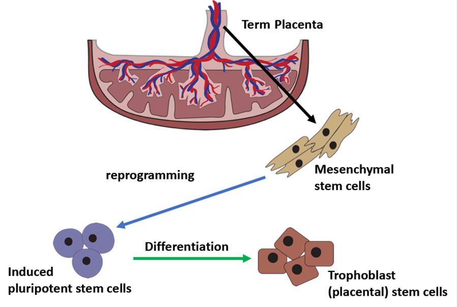
The main functional cell type in the placenta is called “trophoblast” (“tropho” means “to feed”). Different trophoblast subtypes carry out a variety of functions in the placenta: for example, syncytiotrophoblast (STB) are responsible for gas/nutrient exchange, while extravillous trophoblast (EVT) invade the uterus, and establish crosstalk with the maternal immune system to induce tolerance. These different trophoblast subtypes can be isolated from the placenta at term; however, such cells would need to be studied right away and are difficult to manipulate for functional studies. In order to generate trophoblast which are both self-renewing and able to generate the different trophoblast subtypes, we need a method to isolate and culture “trophoblast stem cells” (TSC’s). While such a method has been developed to derive such cells from human embryos and early gestation human placentae, these cells are, by definition, derived from pregnancies whose outcomes are unknown. Our researchers have developed a method by which cells from the placenta at delivery can be reprogrammed first into “induced pluripotent stem cells” (iPSC’s) and subsequently differentiated into TSC’s. These cells can be studied in the dish, not just for detailed analysis of their phenotype, but also for screening of various therapeutic agents to reverse the disease phenotype. In this manner, we are able to model placental diseases that are the basis for many pregnancy complications, including preeclampsia, fetal growth restriction, placenta accreta, and preterm labor.

3D Placenta model.
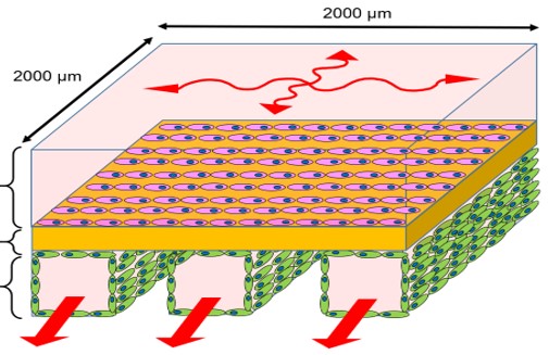
While the majority of studies with cells is done in two-dimensions, organs (including the placenta) exist and function as three-dimensional structures. In particular, the study of the placenta as a barrier through which gas and nutrient exchange occurs is very difficult in two-dimensions. We are therefore collaborating with colleagues in the Nanoengineering Department to develop a “placenta-on-a-chip,” using both primary cells (isolated directly from the placenta) and iPSC-derived cells. This model would allow modeling of the placenta at different gestational ages, thus providing a platform for investigating transport of nutrients, pathogens, and drugs across this barrier.
