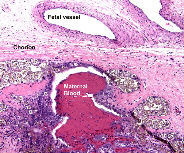
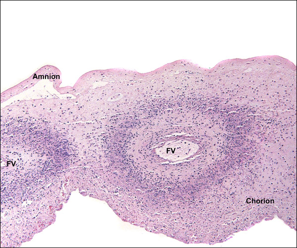
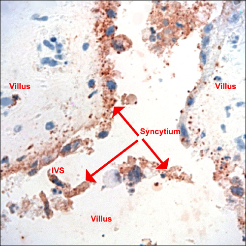
Numerous infectious diseases affect sheep. They have been summarized in the veterinary literature, e.g. by Smith et al. (1972). Among these is brucellosis due to infection with Brucella ovis. It causes mainly epididymitis in rams but may affect the female generative tract and lead to abortion with placental inflammation.
16) Physiological data
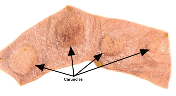
The sheep has been used for numerous physiological studies; many are designed to better understand human prenatal development. Thus, Barron (1951) reviewed in some detail the transfer of oxygen. He and others have studied this transfer at high altitude and compared it with data found at sea level. Liggins (1969) reviewed the endocrine signals for the onset of labor and parturition. Kleemann et al. (2001) showed that progesterone administration in very early pregnancy enhances fetal growth. Alexander (1964) removed up to 84 caruncles from Merino sheep prior to mating in order to assess the impact on placentation. While fertility was not affected, the ewes in which sixty or more caruncles had been removed delivered prematurely and the lambs had reduced birth weights. The caruncle removal was compensated by an increased size of the cotyledons. Worthington et al. (1981) carried out similar reductions with electrocautery. They found a similar effect on fetal growth. Creasy et al. (1972) embolized fetal cotyledons with microspheres between days 96 and 125 and found significant reduction in fetal size. The affected cotyledons showed hyalinization and necrosis with marked reduction in size. This was followed by similar experiments of Duncan et al. (2000). These investigators examined the consequences of interference with placentation on fetal brain development. They identified significant damage to the white matter of the CNS in the growth-restricted lambs. There are many other, similar studies that probe fetal CNS development by restricting placental performance. For instance, Dawes & Mott (1964) altered oxygen supply to fetal lambs and studied their responses. Keunen et al. (1997) found no CNS damage with cord occlusion of up to ten minutes, but Ball et al. (1994) found that seizures took place when there was severe cord occlusion for 90 minutes. Ikeda et al. (1998) sought to explain the CNS lesions after cord occlusion by brain lipid peroxidation. Mallard et al. (1992) found primarily hippocampal lesions after occlusion, while Clapp et al. (1988) found mostly white matter damage after intermittent occlusion of the cord's vessels. Gardner et al. (2002) used sheep placentation to study the effect of periodic cord occlusion. They found that the number of caruncles did not decrease, but their weights decreased and appearances changed.
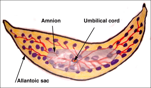
Gilbert et al. (1996) showed that technetium crossed the ovine placenta more rapidly than the smaller sodium ion. Ovine urine production was studied by Ross, et al. (1988) and by many other investigators who are listed by these authors. Ross et al. found in this carefully executed protocol (days after surgical canulations, standing ewes, 4-hour experiment, careful electrolyte measurements) that the amount of allantoic fluid was greater than that of amniotic fluid; that fetal urination is greater into the allantoic sac (through urachus) than the amnionic sac (through urethra); that transmembranous flow probably exists; that lung fluid participates in amnionic fluid composition and swallowing in disposing of it. These are difficult experiments whose interpretation is complicated by the presence of two large fluid-filled sacs in the ovine placenta. They are not directly transferable to human amnionic fluid metabolism for that reason. With Barron we catheterized the urachus in an instrumented sheep that was suspended in a water bath. We found that urine production decreased markedly over a few hours when all urine from the urachus was collected externally. Its osmolality also increased and much fructose was contained in the urine. Urination increased rapidly when water was injected into the allantoic cavity. We conjectured that the water was rapidly absorbed by the allantoic circulation and rehydrated the water-deprived fetus. Matsumoto et al. (2000) ligated the fetal esophagus and urachus and were unable to produce polyhydramnios, as had been their aim. This suggested to them that transmembranous transport must be large.
More recently, Daneshmand et al. (2003) investigated the regulation of amnionic fluid volume in sheep and suggested that vascular endothelial growth factor increases intramembranous absorption by microvesicular transport.
17) Other resources
Cell strains of various sheep species are available from CRES at the Zoological Society of San Diego.
18) Other remarks - What additional Information is needed?
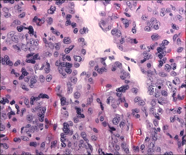
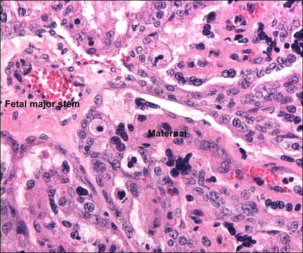
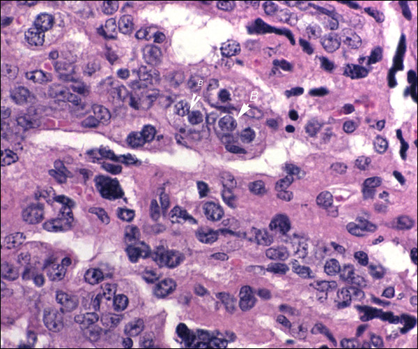
Relatively few noninfectious placental lesions have been described in sheep. More knowledge is also needed on umbilical cords. Mossman (1987) pointed out that more information is needed on the insertion of the cords of multiple gestations. Because there is still some uncertainty as to the true origin of the syncytial giant cells, additional studies are warranted.
Acknowledgement
Most of the animal photographs in these chapters come from the Zoological Society of San Diego. I appreciate also very much the help of the pathologists at the San Diego Zoo.
References
Alexander, G.: Studies of the placenta of the sheep (Ovis aries L.). Placental size. J. Reprod. Fertil. 7:289-305, 1964.
Alexander, G.: Studies on the placenta of the sheep (L.). Effect of surgical reduction in the number of caruncles. J. Reprod. Fertil. 7:307-322, 1964.
Arvy, L. and Pilleri, G. The Cordon Ombilical (Funis umbilicalis). Verlag Hirnanatom. Instit., Ostermundigen, Switzerland, 1976.
Assheton, R.: VI. The morphology of the ungulate placenta, particularly the development of that organ in the sheep, and notes upon the placenta of the elephant and hyrax. Phil. Trans. Roy. Soc. London, Series B 198:143-220, 1906.
Ball, R.H., Parer, J.T., Caldwell, L.E. and Johnson, J.: Regional blood flow and metabolism in ovine fetuses during severe cord occlusion. Amer. J. Obstet. Gynecol. 171:1549-1555, 1994.
Barron, D.H.: Some aspects of the transfer of oxygen across the syndesmochorial placenta of the sheep. The Yale J. Biol. Med. 24:169-190, 1951.
Bautzmann, H. and Schroder, R.: Vergleichende Studien uber Bau und Funktion des Amnions. Das Amnion der Säuger am Beispiel des Schafes (Ovis aries). Z. Zellforsch. 43:48-63, 1955.
Bourne, G.L.: The Human Amnion and Chorion. Lloyd-Luke, London, 1962.
Bunch, T.D., Foote, W.C. and Spillett, J.J.: Sheep-goat hybrid karyotypes. Theriogenology 6:379-385, 1976.
Clapp, J.F., Peress, N.S., Wesley, M. and Mann, L.I.: Brain damage after intermittent partial cord occlusion in the chronically instrumented fetal lamb. Amer. J. Obstet. Gynecol. 159:504-509, 1988.
Creasy, R.K., Barrett, C.T., Swiet, M.de, Kahanpää, K.V. and Rudolph, A.M.: Experimental intrauterine growth retardation in the sheep. Amer. J. Obstet. Gynecol. 112:566-573, 1972.
Daneshmand, S.S., Cheung, C.Y. and Brace, R.A.: Regulation of amniotic fluid volume by intramembranous absortion in sheep: Role of passive permeability and vascular endothelial growth factor. Amer. J. Obstet. Gynecol. 188:786-793, 2003.
Davies, J.: Correlated anatomical and histochemical studies on the mesonephros and placenta of the sheep. Amer. J. Anat. 91:263-299, 1952.
Davies, J. and Wimsatt, W.A.: Observations on the fine structure of the sheep placenta. Acta anat. 65:182-223, 1966.
Dawes, G.S. and Mott, J.C.: Changes in O2 distribution and consumption in foetal lambs with variation in umbilical blood flow. J. Physiol. 170:524-540, 1964.
Dent, J., McGovern, P.T. and Hancock, J.L.: Immunological implications of ultrastructural studies of goat x sheep hybrid placentae. Nature 231:115-117, 1971.
Duncan, J.R., Cock, M.L., Harding, R. and Rees, S.M.: Relation between damage to the placenta and the fetal brain after late-gestation placental embolization and fetal growth restriction in sheep. Amer. J. Obstet. Gynecol. 183:1013-1022, 2000.
Gardner , D.S., Ward, J.W., Giussani, D.A. and Fowden, A.L.: The effect of a reversible period of adverse intrauterine conditions during late gestation on fetal and placental weight and placentome distribution in sheep. Placenta 23:459-466, 2002.
Gardner, D.S., Ward, J.W., Giussani, D.A. and Fowden, A.L.: The effect of a reversible period of adverse intrauterine conditions during late gestation on fetal and placental weight and placentome distribution in sheep. Placenta 23:459-466, 2002.
Gilbert, W.M., Newman, P.S., Eby-Wilkens, E. and Brace, R.A.: Technetium Tc 99m rapidly crosses the ovine placenta and intramembranous pathway. Amer. J. Obstet. Gynecol. 175:1557-1562, 1996.
Gray, A.P.: Mammalian Hybrids. Second edition. A Check-List with Bibliography. Commonwealth Agricultural Bureaux, Farnham Royal, Slough, UK, 1972.
Houston, F., Foster, J.D., Chong, A., Hunter, N. and Bostock, C.J.: Transmission of BSE by blood transfusion in sheep. Lancet 356:999-1000 (and 955-956 for Comments), 2000.
Hyde, S.R. and Benirschke, K.: Gestational psittacosis: Case report and literature review. Modern Pathol. 10:602-607, 1997.
Ikeda, T., Murata, Y., Quilligan, E.J., Parer, J.T., Doi, S. and Park, S-D.: Brain lipid peroxidation and antioxidant levels in fetal lambs 72 hours after asphyxia by partial umbilical cord occlusion. Amer. J. Obstet. Gynecol. 178:474-478, 1998.
Johnson, G.A., Burghardt, R.C., Joyce, M.M., Spencer, T.E., Bazer, F.W., Pfarrer, C. and Gray , C.A. : Osteopontin expression in uterine stroma indicates a decidualization-like differentiation during ovine pregnancy. Biol. Reprod. 68:1951-1958, 2003.
Keisler, D.H.: Sheep and Goats. In, Encyclopedia of Reproduction. E. Knobil and J.D. Neill, eds. Vol. 4, pp. 479-492. Academic Press, San Diego, 1999.
Keunen, H., Blanco, C.E., Reempts, J.L.H.van and Hasaart, T.H.M.: Absence of neuronal damage after umbilical cord occlusion of 10, 15, and 20 minutes in midgestation fetal sheep. Amer. J. Obstet. Gynecol. 176:515-520, 1997.
King, G.J., Atkinson, B.A. and Robertson, H.A.: Implantation and early placentation in domestic ungulates. J. Reprod. Fertil. Suppl. 31:17-30, 1982.
Kleemann, D.O., Walker, S.K., Hartwich, K.M., Fong, L., Seamark, R.F., Robinson, J.S. and Owens, J.A.: Fetoplacental growth in sheep administered progesterone during the first three days of pregnancy. Placenta 22:14-23, 2001.
Lawn, A.M., Chiquoine, A.D. and Amoroso, E.C.: The development of the placenta in the sheep and goat: An electron microscopic study. J. Anat. 105:557-578, 1969.
Lee, C.S., Gogolin-Ewens, K. and Brandon, M.R.: Comparative studies on the distribution of binucleate cells in the placentae of deer and cow, using monoclonal antibody, SBU-3. J. Anat. 147:163-179, 1986.
Liggins, G.C.: The foetal role in the initiation of parturition in the ewe. CIBA Fndt. Symp. On Foetal Anatomy. G.E.W. Wolstenholm & M. O'Connor, eds. Pp. 218-231, 1969.
Long, S.E. and Williams, C.V.: Frequency of chromosomal abnormalities in early embryos of the domestic sheep (Ovis aries). J. Reprod. Fertil. 58:197-201, 1980.
Ludwig, K.S.: Zur Feinstruktur der materno-fetalen Verbindung im Placentom des Schafes (Ovis aries L.). Experientia 18:212-213, 1962.
Ludwig, K.S.: Zur vergleichenden Histologie des Allantochorion. Rev. Suisse Zool. 75:819-831, 1968.
Makowski, E.L.: Maternal and fetal vascular nets in placentas of sheep and goats. Amer. J. Obstet. Gynecol. 100:283-288, 1968.
Mallard, E.C., Gunn, A.J., Williams, C.E., Johnston, B.M. and Gluckman, P.D.: Transient umbilical cord occlusion causes hippocampal damage in the fetal sheep. Amer. J. Obstet. Gynecol. 167:1423-1430, 1992.
Matsumoto, L.C., Cheung, C.Y. and Brace, R.A.: Effect of esophageal ligation on amniotic fluid volume and urinary flow rate in fetal sheep. Amer. J. Obstet. Gynecol. 182:699-705, 2000.
Miyazaki, H., Imai, M., Hirayama, T., Saburi, S., Tanaka, M., Maruyama, M., Matsuo, G., Meguro, H., Nishibashi, K., Inoue, F., Djiane, J., Gertler, A., Tachi, S., Imakawa, K. and Tachi, C.: Establishment of feeder-independent cloned caprine trophoblast cell line which expresses placental lactogen and interferon tau. Placenta 23:613-630, 2002.
Mossman, H.W.: Vertebrate Fetal Membranes. MacMillan, Houndmills, 1987.
Naaktgeboren, C. and Zwillenberg, H.H.L.: Untersuchungen uber die Auswuchse am Amnion und an der Nabelschnur bei Walen und Huftieren, mit besonderer Berucksichtigung des europäischen Hausrindes. Acta morphol. Neerlando-Scand. 4:31-60, 1961.
Nowak, R.M.: Walker's Mammals of the World. 6th ed. The Johns Hopkins Press, Baltimore, 1999.
Osgerby, J.C., Gadd, T.S. and Wathes , D.C. : The effects of maternal nutrition and body condition on placental and foetal growth in the ewe. Placenta 24:236-247, 2003.
Puschmann, W.: Zootierhaltung. Vol. 2, Säugetiere. VEB Deutscher Landwirtschaftsverlag Berlin, 1989.
Reynolds, S.R.M.: The proportion of Wharton's jelly in the umbilical cord in relation to distention of the umbilical arteries and vein, with observations on the folds of Hoboken. Anat. Rec. 119:365-377, 1952.
Ross, M.G., Ervin, M.G., Rappaport, V.J., Youssef, A., Leake, R.D. and Fisher, D.A.: Ovine fetal urine contribution to amniotic and allantoic compartments. Biol. Neonat. 53:98-104, 1988.
Silverstein, A.M., Prendergast, R.A. and Kraner, K.L.: Homograft rejection in the fetal lamb: The role of circulating antibody. Science 142:1172-1173, 1963.
Silverstein, A.M., Thorbecke, G.J., Kraner, K.L. and Lukes, R.J.: Fetal response to antigenic stimulus. III. ?-globulin production in normal and stimulated lambs. J. Immunol. 91:384-395, 1963.
Smith, H.A., Jones, T.C. and Hunt, R.D.: Veterinary Pathology. Lea & Febiger, Philadelphia, 1972.
Ward, J.W., Wooding, F.B.P. and Fowden, A.L.: The effect of cortisol on the binucleate cell population in the ovine placenta during late gestation. Placenta 23:451-458, 2002.
Wilsmore, A.J., Izzard, K.A., Wilsmore, B.C. and Dagnall, G.I.R.: Breeding performance of sheep infected with Chlamydia psittaci (ovis) during their preceding pregnancy. Vet. Rec. 130:68-70, 1990.
Wimsatt, W.A.: New histological observations on the placenta of the sheep. Amer. J. Anat. 87:391-458, 1950.
Wooding, F.B.P.: Role of binucleate cells in fetomaternal cell fusion at implantation in the sheep. Amer. J. Anat. 170:233-250, 1950.
Wooding, F.B.P.: Frequency and localization of binuclear cells in the placentome of ruminants. Placenta 4:527-540, 1983.
Wooding, F.B.P., Flint, A.P.F., Heap, R.B. and Hobbs, T.: Autoradiographic evidence for migration and fusion of cells in the sheep placenta: Resolution of a problem in placenta classification. Cell Biol. Internat. Reports 5:821-827, 1981. See also: Cell Biol. Int. Rep. 5:821-827, 1981.
See also: Cell Biol. Int. Rep. 5:821-827, 1981.Wooding, F.B., Flint , A.P., Heap, R.B., Morgan, G., Buttle, H.L. and Young, I.R.: Control of binucleate cell migration in the placenta of sheep and goat. J. Reprod. Fertil. 76:499-512, 1986.
Wooding, F.B., Flint, A.P., Heap, R.B., Morgan, G., Buttle, H.L. and Young, I.R.: Control of binucleate cell migration in the placenta of sheep and goat. J. Reprod. Fertil. 76:499-512, 1986.
Worthington, D., Piercy, W.N. and Smith, B.T.: Effects of reduction of placental size in sheep. Obstet. Gynecol. 58:215-221, 1981.
|
